


There is a trade-off between completeness and readability in plain text.
The more detail we add to a paragraph, the more difficult it is for the reader to get through it.
In research methods, we have a lot of details. To make sure what you release out there can be reproduced, you need to add as much detail as possible. But the more detail you add, the less it feels like you are writing in English. And, when authoring methods sections myself, time and time again I compromised on readability in favour of completeness.
When reading a paper, processing those methods sections is no joke. I find my eyes running up and down the paragraph, struggling to understand the relationships and piece everything together. It's a mental workout to grasp what happened in the lab and how.
Consider this methods section example. Notice how long it takes you to understand it, how your eyes move across the text, and which details you notice first.
In this study, transfection was performed on HEK293 cells, which had been cultured to 70-80% confluency in a controlled environment at 37°C and 5% CO2. For this experiment, SiRNA was used, with Lipofectamine 2000 being employed in a 1:1 ratio. Both of these components were diluted separately in 100 µl of Opti-MEM. The mixture of siRNA and Lipofectamine, after being gently combined, was allowed to stand at room temperature (approximately 22°C) for 40 minutes to facilitate the formation of siRNA-Lipofectamine complexes. Meanwhile, the cell viability was assessed using a 0.4% trypan blue dye, using fluorescence microscopy at 560 nm excitation and 590 nm emission, establishing baseline health prior to transfection. The siRNA-Lipofectamine complexes were then introduced into the cells in a dropwise manner, ensuring even distribution. This introduction was followed by a 6-hour incubation period at 37°C, after which the medium was replaced with DMEM enriched with 10% FBS and 1% penicillin-streptomycin, to nourish and protect the cells post-transfection.I usually start by fishing out the actions – and anchor there. Then, I look for the relevant parameters characterising the actions, e.g. at 37°C and 5% CO2. And, finally, move onto auxiliary information, like observations of the cells or vendor information.
Well, at least that’s what I try to do. Sometimes reading the extra fluff is easier because it feels more like a narration. So, my brain might take the path of least resistance and go to those bits of information first instead.
The extra level of complexity comes from the use of passive forms, e.g., siRNA was used, and from separating relevant information from the actual step, e.g., the 1:1 ratio mentioned before the mixing step. Let’s take our example paragraph again and highlight the main lab steps.
In this study, transfection was performed on HEK293 cells, which had been cultured to 70-80% confluency in a controlled environment at 37°C and 5% CO2. For this experiment, SiRNA was used, with Lipofectamine 2000 being employed in a 1:1 ratio. Both of these components were diluted separately in 100 µl of Opti-MEM. The mixture of siRNA and Lipofectamine, after being gently combined, was allowed to stand at room temperature (approximately 22°C) for 40 minutes to facilitate the formation of siRNA-Lipofectamine complexes. Meanwhile, the cell viability was assessed using a 0.4% trypan blue dye, using fluorescence microscopy at 560 nm excitation and 590 nm emission, establishing baseline health prior to transfection. The siRNA-Lipofectamine complexes were then introduced into the cells in a dropwise manner, ensuring even distribution. This introduction was followed by a 6-hour incubation period at 37°C, after which the medium was replaced with DMEM enriched with 10% FBS and 1% penicillin-streptomycin, to nourish and protect the cells post-transfection.Now, let’s make the sentences active and reorganise the information.
We'll also start each step on a new line for easier reading.
Prior to transfection, we cultured HEK293 cells to 70-80% confluency in a controlled environment at 37°C and 5% CO2.
We diluted siRNA and Lipofectamine 2000 separately in 100 µl of Opti-MEM.
We gently combined mixture of siRNA and Lipofectamine at 1:1 ratio.
We incubated the mixture at room temperature (approximately 22°C) for 40 minutes to facilitate the formation of siRNA-Lipofectamine complexes.
Meanwhile, we assessed cell viability using a 0.4% trypan blue dye, using fluorescence microscopy at 560 nm excitation and 590 nm emission to establish baseline health prior to transfection.
We then added siRNA-Lipofectamine complexes to the cells in a dropwise manner, ensuring even distribution.
We incubated the mixture for 6-hour at 37°C.
We replaced the medium with DMEM enriched with 10% FBS and 1% penicillin-streptomycin, to nourish and protect the cells post-transfection.
Okay, this is already much more readable. Skimming through this methods section, I can quickly get the gist. So even if I'm searching for a specific detail, I will roughly know where to look for it.
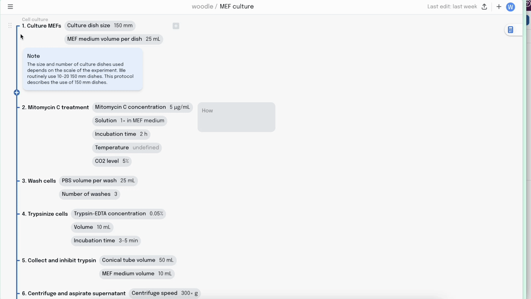
But how do we find those specific details? First, we probably formulate a request in our minds. For example, what ratio was used to mix siRNA and Lipofectamine? And then we look for it. So what if each step had a dedicated section for these key details, clearly labeled?

This feels a bit more queryable. You might notice some details are missing now, making the method seem dry. In every good method, there are free-text notes that provide additional context, like why a step is important, or how it is run. Let’s add a special field for those.

Now, it’s easier to navigate through the different parts and zoom into the relevant information. But there is one more piece missing – the “while”.
We all know there are plenty of wait times in the lab – and it always feels good to use those efficiently. But plain text lacks the means to show what steps can be parallelised. What if we visually connect steps that belong to the same process and indicate when different processes overlap?

We called this view a Tube Map [thought it looked familiar?].
Here, all the most important information [the lab actions] is discoverable when traveling along the Tube Lines. Once at the station, we can dig into more detail by sliding our eyes to the right. The less core the information is, the longer the travel distance from the station will be.
Now, when reading, we can layer the description on in any order or quickly zoom into just what we are looking for. And when designing our own method, we can optimise the parallelisation and add as much detail as we want – without sacrificing readability.

There's a catch, though. It does take substantially longer to categorise each detail and organise the information when creating a new method. And a couple of years ago, this would have been the end of the story. No one has the time to do all this legwork just to make the methods section look better.
But in our age of LLMs, the task of formatting plain text into a standardised representation becomes a piece of cake. And even though there are occasional mistakes here and there, the quality of the translation between plain text and the tube map format is pretty damn good. Check it out.
Honestly, this is the only way I read papers now.
And if you want to give it a go yourself, sign up here – and we’ll give you access to our alpha tool.